Acoustic Neuroma
Acoustic neuroma is a noncancerous growth which can develop on the eighth cranial nerve. Also termed as the vestibulocochlear nerve, it is known to connect the inner ear with the brain, and it consists of two different parts. While one part is responsible for the transmission of sound, the other part helps to send balance information from the inner ear to the brain.
Acoustic neuromas, which may also be termed vestibular schwannomas, or neurilemmomas, generally grow slowly over a period of years. They may not actually invade the brain, but they can push on it as they continue to grow. Larger tumors can press on the nearby cranial nerves that control the muscle of facial expression as well as sensation. If the tumors get large enough to press on the brain stem or the cerebellum, then they can be quite deadly.
Symptoms
Signs and symptoms of acoustic neuroma are mostly subtle, and it can take several years to fully develop. They generally arise from the tumor’s effects on the hearing and balance nerves. Pressure caused by the tumor on the adjacent nerves that control the facial muscles and sensation, brain structures, or nearby blood structures can also cause problems.
As the tumor continues to grow, it might be more likely to cause more noticeable or severe signs and symptoms. Some of the common signs and symptoms of an acoustic neuroma can include the following:
- Hearing loss, which is usually gradual. However, in some cases, it may be sudden. It might also occur on only one side or more pronounced on one side.
- Ringing in the affected ear which is also termed as tinnitus
- Facial numbness and very rarely, weakness or loss of muscle movement
- Unsteadiness, or loss of balance
- Dizziness, which can also be termed as vertigo
In some rare cases, an acoustic neuroma can even grow large enough and compress the brainstem, which may become life-threatening.
If you notice significant hearing loss in one ear, ringing in your ear, or trouble with your balance, then you should consider seeing your doctor soon.
Early diagnosis of an acoustic neuroma might help in keeping the tumor from growing large enough to cause any serious consequences, such as total hearing loss or a life-threatening buildup of fluid within the skull.
Causes
Two types of acoustic neuroma exist: a sporadic form and a form associated with a syndrome which is known as neurofibromatosis type II (NF2). NF2 is an inherited disorder that is characterized by the growth of noncancerous tumors in one’s nervous system. Acoustic neuromas are also known to be the most common of these tumors and they generally occur in both ears by the age of 30.
NF2 is a rare disorder, and it accounts for only around 5 percent of acoustic neuromas. This means the vast majority of the cases are the sporadic form. Doctors are however uncertain what exactly leads to this sporadic form. One of the known risk factors for this condition is exposure to high doses of radiation, especially to one’s head or neck.
Diagnosis
In the early stages, it is generally difficult to diagnose acoustic neuroma, since signs and symptoms might be subtle and can develop over time gradually. Some of the common symptoms include hearing loss, which is associated with several middle and inner ear problems.
After you ask questions about your symptoms, your doctor will be conducting an ear exam. Your doctor might order the following tests.
Hearing test (audiometry)
Imaging
Treatment
There are three main courses of treatment which are used for acoustic neuroma:
- Observation
- Surgery
- Radiation therapy
Observation
Surgery
Surgery for acoustic neuromas involves removing the entire or part of the tumor.
Three main surgical approaches exist, for the removal of an acoustic neuroma:
Translabyrinthine
Translabyrinthine involves making an incision behind the ear and then removing the bone behind the ear along with some of the middle ear. This procedure is generally used for tumors which are larger than 3 centimeters. The upside of this approach is that it can allow the surgeon to see an important cranial nerve or the facial nerve quite clearly before he/she removes the tumor. The downside to this technique is that it causes permanent hearing loss.
Retrosigmoid/sub-occipital
Retrosigmoid/sub-occipital method involves exposing the back of the tumor by opening the skull near the back of one’s head. This approach can be used in order to remove tumors of any size and it also offers the possibility of preserving one’s hearing ability.
Middle fossa
Radiation therapy
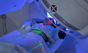
Radiation therapy might be recommended in certain cases for acoustic neuromas. Due to state-of-the-art delivery techniques, it is possible to send high doses of radiation to the tumor while at the same time, limiting exposure and damage to any surrounding tissue.
The tumor’s growth might slow or stop or it might even shrink, however, the radiation doesn’t remove the tumor completely.
Complications
An acoustic neuroma can lead to a variety of permanent complications, which can include the following:
- Hearing loss
- Ringing in the ear
- Facial numbness and/.or weakness
- Difficulties with balance
Large tumors may press on your brainstem, which can prevent the normal flow of fluid between the brain and the spinal cord. In this case, fluid may build up in your head, which can increase the pressure inside the skull.


