Best Doctors in India for AVM Brain Treatment
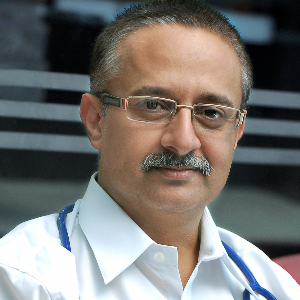
- Cardiac Surgeon, Gurugram, India
- Over 40 years’ experience
- Medanta-The Medicity, Gurgaon
Profile Highlights:
- Dr. Naresh Trehan is one of the most highly skilled and globally recognized cardiothoracic and vascular surgeons.
- His experience encompasses 40+ years and is considered one of the most successful and accomplished cardiac surgeons in the country.
- Dr. Naresh Trehan specializes in cardiovascular surgery, cardiothoracic surgery, Heart Transplant, and Minimally Invasive Cardiac Surgery and has performed more than 48,000 successful open-heart surgeries to date.
- He started his career as a Cardiac Surgeon at New York University Medical Centre where he performed over 3000 coronary artery surgeries and returned to India in 1988 and founded the Escorts Heart Institute and Research Center in New Delhi.
- Dr. Trehan is the Founder Chairman of Medanta- The Medicity in Gurugram, a premier multi-specialty hospital with state-of-the-art infrastructure and equipped with the latest and advanced technologies.
- Top Pulmonologist and Respiratory Medicine specialist | Apollo Hospital, New Delhi, India
- 48+ Years Experience
- Indraprastha Apollo Hospital, New Delhi
Profile Highlights:
- Dr. M.S. Kanwar is a distinguished pulmonologist with an impressive 46-year career, renowned both in India and internationally for his expertise in COVID-19, sleep medicine, and pulmonary care.
- Dr. Kanwar’s pioneering spirit extends to sleep medicine, where he made history in 1995 by establishing the largest and most advanced sleep lab in India at Indraprastha Apollo Hospital in New Delhi. His work in this area has set a high standard for sleep disorder management in the country.
- His other areas of expertise include asthma, COPD, lung infections, COVID-19, lung fibrosis, ILD, tuberculosis, lung cancer, sarcoidosis, lung transplants, ICU-related diseases, sepsis, and mechanical ventilation. He is skilled in advanced procedures such as bronchoscopy, EBUS (Endobronchial Ultrasound), and medical thoracoscopy.
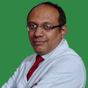
- Pediatric Cardiac Surgeon, New Delhi, India
- Over 30 years’ experience
- Marengo Asia Hospital, Faridabad
Profile Highlights:
- Dr. Rajesh Sharma is one of the most experienced and widely acclaimed Cardiothoracic and Vascular surgeons specializing in pediatric cardiac surgery in India.
- He provides treatment for both adult and pediatric heart diseases and disorders and focuses primarily on complex congenital heart diseases.
- Dr. Sharma’s expertise lies in infant and Neonatal cardiac surgery, surgery for transposition of the great arteries, Fontan circulation, and acquired heart diseases.
- His experiences encompass over 3 decades during which he has performed over 20,000 heart surgeries including congenital heart disorders as well as acquired heart defects.
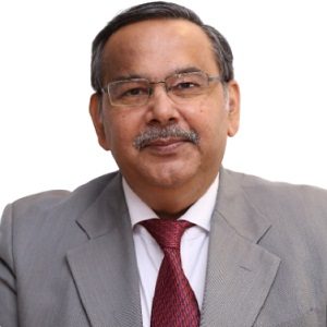
- Hematologist & BMT Specialist, Gurugram, India
- Over 20 years’ experience
- Fortis Memorial Research Institute
Profile Highlights:
- Dr. Rahul Bhargava is a leading Hematologist and BMT specialist currently working at Fortis Memorial Research Institute, Gurugram.
- Dr. Bhargava has an experience of more than 20 years in Hematology, Pediatric Hemato Oncology, and BMT during which he has performed numerous BMTs with successful outcomes.
- He primarily specializes in Haploidentical and unrelated match transplants and has performed over 1500 transplants till date.
- In 2016, he became the first Indian doctor to perform and popularize BMT for Multiple Sclerosis.
- Top Rheumatologist | Apollo Hospital, New Delhi, India
- 30+ Years Experience
- Indraprastha Apollo Hospital, New Delhi
Profile Highlights:
- Dr. Sundeep Kumar Upadhyaya is one of the best Rheumatologists in India, working at Indraprastha Apollo Hospitals, New Delhi.
- He has over 30 years of experience in modern medicine, spondylitis, rheumatology, arthritis, and other connective tissue disorder treatment.
- Dr. Upadhyaya has also been a crucial part of various research.
- He was one of the first rheumatology experts in India to introduce bioagents to treat rheumatoid arthritis, inflammatory spondylitis, and ankylosing spondylitis.
- Dr. Upadhaya provides training for PG students in Apollo Hospital, New Delhi.
- Top Rheumatologist | Apollo Hospital, New Delhi, India
- 35+ Years Experience
- Indraprastha Apollo Hospital, New Delhi
Profile Highlights:
- With over 40 years of distinguished experience in Rheumatology, Dr. Prof. Rohini Handa is a leading Rheumatologist based at Indraprastha Apollo Hospitals, New Delhi. A celebrated figure in his field, he has a robust background that combines deep clinical expertise with significant academic and leadership roles.
- Dr. Rohini Handa is renowned for his expertise in managing a range of rheumatologic conditions, including Rheumatoid Arthritis, Myositis (muscle inflammation), Sjogren’s Syndrome (a condition leading to dry eyes and mouth), and Multiminicore Disease (a muscle disorder).
- He excels in performing key diagnostic and therapeutic procedures such as analyzing synovial fluid (the fluid within joints), conducting joint injections and aspirations (to remove excess fluid from joints), and administering cortisone injections (steroid treatments to alleviate inflammation).
Best Hospitals in India for AVM Brain Treatment
- City: Bengaluru, India
Hospital Highlights:
- Fortis Hospital Bannerghatta, Bengaluru was established in 2006.
- The hospital is a 276 bedded multi-specialty tertiary care facility.
- The hospital specializes in cutting-edge medical technology and dedicated patient care services.
- The hospital is equipped with state-of-the-art technologies like trans-radial angioplasty, trans-abdominal cardiac surgery, and computerized TKR navigation surgery.
- The hospital provides specialty medical services in cardiology, cardiac surgery, orthopedics, neurology, neuro-surgery, GI, and Minimal Access Surgery (MAS).
- City: Chennai, India
Hospital Highlights:
- Fortis Malar was established in 1992 and was formerly known as Malar Hospital.
- The hospital specializes in cutting-edge medical technology and dedicated patient care services.
- The hospital is multi-specialty, tertiary care facility with 180 beds.
- The hospital offers comprehensive medical care in specialties such as cardiology, cardio-thoracic surgery, neurology, neurosurgery, orthopedics, nephrology, gynecology, gastroenterology, urology, pediatrics, and diabetes.
- City: New Delhi, India
Hospital Highlights:
- Established in 1996, Pushpawati Singhania Research Institute is one of the top hospitals in the NCR region, as well as one of the top facilities in India for gastroenterology. The hospital is one of South Asia’s first institutes in medical and surgical treatment for diseases related to digestion.
- The hospital is equipped with state-of-the art facilities coupled with the latest equipment as well as renowned consultants from various parts of India as well as other parts of the world.
- City: New Delhi, India
Hospital Highlights:
- State-of-the-art technology and devoted healthcare professionals have been brought together under one roof at Venkateshwar Hospital to provide genuine medical care. The hospital’s professionals work together as a team to deliver the best possible treatment to their patients, using the most sophisticated equipment and information technology.
- Venkateshwar Hospital’s mission is to attain global excellence in healthcare by employing evidence-based, ethical clinical practices and cutting-edge technology by a team of highly skilled experts.
- City: New Delhi, India
Hospital Highlights:
- Sir Ganga Ram Hospital, New Delhi is known to provide the latest medical procedures with the latest technology in all of its units.
- The hospital has a team of reputed doctors, nurses, and healthcare professionals that ensure that patients receive quality care at affordable costs.
- Staffed with a team of highly qualified doctors, dedicated nurses, and paramedical and non-medical staff, the hospital aims to lead in healthcare delivery, medical education, training, and research.
- As per the vision of the founder, the hospital also provides free treatment to the economically weaker sections of society.
- Sir Ganga Ram Hospital also provides training to young doctors under the Diplomate in National Board(DNB) program. The DNB program at the hospital was started in 1984 and it is known for currently running the maximum number of DNB specialties in the country. It also has the distinction of having the first bone bank in India.
- City: Kerala, India
Hospital Highlights:
- Established in 2019, Apollo Adlux Hospital is the first Apollo Hospital in Kerala and the 73rd hospital owned by Apollo Group in India. With the state’s most advanced, comprehensive healthcare infrastructure and cutting-edge technologies, Apollo Adlux Hospital stands as an example of medical excellence in Kerala.
- With over 34 multi-specialty departments, the hospital believes in providing the best quality treatment to its patients at affordable rates, ensuring comfort at their difficult times.
- The 300-bed hospital is managed by a team of highly qualified and experienced experts who delivers exceptional hospitality to their patients and treats them with great compassion.
- With its affiliation with the Apollo Hospitals Group, the hospital aims in providing patients with top-notch healthcare services while also serving communities in Kerala.
- The hospital has good railway and road connections, and is conveniently close to Cochin International Airport.
- City: Gurugram, India
Hospital Highlights:
- Situated near DLF Cyber City, Gurugram, Narayana Superspecialty Hospital is one of the top medical facilities in the Delhi NCR region, catering to the needs of the people. Known for its commitment to quality medical care and patient service, the hospital is a state-of-the-art facility with planned and well-equipped sections, which includes a spacious OPD area as well as comfortable patient rooms.
- It is the closest super-specialty hospital from Indira Gandhi International Airport towards Gurugram, and also the nearest super specialty hospital from DLF Cyber City. It is also close to major residential areas in Gurugram.
- It is part of the renowned Narayana Health Group. Established in 2000, by Dr. Devi Shetty, a renowned cardiac surgeon, it has grown to be one fo India’s leading healthcare groups.
- City: Noida, India
Hospital Highlights:
- Fortis Hospital, Noida, stands as one of the oldest and most trusted healthcare institutions in the region, setting a benchmark for comprehensive medical care.
- As the second mega hub hospital in the Fortis Healthcare Group, Fortis Hospital, Noida, upholds a legacy of trust among more than 1.2 million patients. By integrating top-tier professionals with cutting-edge technology, the hospital delivers superior treatment across various medical disciplines.
- Specializing in advanced Neurosciences, Orthopedics, Kidney and Liver Transplant Programmes, Fortis Hospital, Noida has successfully performed over 1,500 transplants, solidifying its reputation as a leader in specialized medical interventions.
WHAT IS AN AVM BRAIN?
What causes Brain AVM?
The primary cause of AVM is still unclear. Some people have brain AVM by birth while some others develop the same later. A notable point is the passing down of this malformation amongst families, genetically.
Occurrence rate of AVMs
Brain AVMs occur very rarely and their occurrence rate is around 1% of the population. However, you might observe male predilection for AVM of the brain.
Where AVMs occur
Although an AVM may develop anywhere within the body, the most common sites are the brain and spine. However, brain AVM is rare and affects only a small chunk of the human population.
Symptoms of Brain AVMs
You may not notice any symptoms of brain AVM till it ruptures. Rupturing of AVM of the brain may result in bleeding within the brain which is termed hemorrhage. A majority of the patients with brain AVM experience hemorrhage as the first sign. Other symptoms of brain AVM include:
- Constant pain in any specific area of the head
- Problems with speech
- Progressive loss of neurological function
- Numbness or paralysis
- Dizziness
- A problem in understanding language
- Nausea and vomiting
- A problem in performing certain tasks that require planning
- Back pain
- Loss of coordination
- Seizures
- Muscle weakness in a specific part of the body
- Loss of consciousness
- Weak lower limbs
- Confusion
- Vision loss
- Serious unsteadiness
- Hallucinations
- Inability to understand what others are saying
- Loss of control on eye movements
- Tingling sensations
Causes of bleeding AVM
The abnormal blood vessels in brain AVM are weak. Hence, they direct blood in a direction against the tissues of the brain. As there is dilation of the abnormal blood vessels with time, they tend to burst. A brain AVM ruptures and bleeds because of the blood flow in a high pressure coming from the arteries.
Chances of AVM bleeding
A brain AVM generally develops between the age of 10 years to 40 years. On average, the chances of bleeding of brain AVM is between 1% to 3% per year. If it bleeds for the first time, the patients are at risk of recurrent bleeding within a short period. Teenagers and the young age group people are at a high risk of experiencing recurrent bleeding.
When to visit the doctor?
If you notice any signs or symptoms of brain AVM, you must visit the doctor immediately. These cases require emergency medical attention otherwise they may bleed profusely & prove to be fatal.
Stages of brain AVM
- Stage 1 of Quiescence: AVM brain malformation is quiet in this stage. The skin surface above the AVM is red and feels a little warm.
- Stage 2 of Expansion: The malformation increases in size to become larger and it is easy to feel the pulse within the malformation.
- Stage 3 of Destruction: The brain AVM begins to bleed and pain.
- Stage 4 of Decompensation: This stage may turn out to be fatal as heart failure occurs in most cases.
Can AVM bleeding prove fatal?
Around 10% to 15% of deaths occur after each AVM brain bleeding. Some patients experience permanent brain damage that accounts for around 30% of the total cases. Every bleed contributes to the damage of normal tissues within the brain that may alter the normal brain functions temporarily or permanently.
Types of Brain AVM
There are 5 different types of brain AVM. These types are listed as under:
- True AVM: This is the most common type of malformation in which the abnormal blood vessels tangle. However, there is no interfering normal tissue of the brain.
- Venous malformation: This malformation involves only the abnormal veins.
- Dural fistula: Dura mater, the covering of the brain, often gets involved in an abnormal connection with the blood vessels. This results in the formation of a dural fistula. There are three types of dural fistulas namely transverse- sigmoid sinus dural fistula, dural carotid-cavernous sinus fistula, and sagittal sinus-scalp dural fistula.
- Cryptic or occult AVM or cavernous malformations: This type of vascular brain malformation doesn’t draw away large quantities of blood. Such malformations may cause seizures and bleed.
- Hemangioma: Abnormal blood vessels at the brain or skin surface are termed hemangioma.
Risk factors of Brain AVM
Males are at a higher risk of developing brain AVM. Apart from this, families with a history of brain AVM have generic passing down of the same.
Complications of Brain AVM
The complications of brain AVM include stroke, speech or movement problems, low quality of life, developmental delays (in children), and numbness in specific parts of the body. Other complications associated may be:
- Weaker blood vessels: An AVM brain exerts pressure on the weaker blood vessels. This may result in an aneurysm or a bulge in the walls of the blood vessels that may rupture later.
- Hemorrhage: AVM causes the walls of the affected veins and arteries to weaken which results in bleeding within the brain or hemorrhage. While hemorrhage goes undetected in some people, some others experience episodes that may be life-threatening.
- Brain damage: In many cases, AVM increases in size to constrict some portions of the brain. Because of this, protective fluids are unable to circulate freely and instead collect at a place. This may cause brain tissue to get pushed against the skull resulting in brain damage.
- Reduced oxygen supply: When brain AVM develops, blood circulates directly to the veins & the arteries. Because of this, surrounding tissues are unable to absorb oxygen from the blood, and these tissues weaken or die.
How Brain AVMs are diagnosed
Your doctor will review all your symptoms and perform a thorough examination. For forming a diagnosis, he or she might ask you to undergo certain tests. These tests are usually performed by neuroradiologists. They are professionally trained to conduct imaging tests for finding the cause and the diagnosis of the condition.
Tests
- CT scan: A Computerized Tomography scan creates a cross-sectional radiographic image of the brain through an x-ray series. Sometimes, doctors perform angiograms which involves injecting a dye. This dye is injected into the veins through a tube so that minute details can be seen.
- Cerebral arteriography: Also called cerebral angiography, it helps to accurately diagnose AVM. It reveals not only the characteristics of arteries that supply AVM and the draining veins but also the exact location. These details are essential for the diagnosis and treatment of the condition. Your doctor will insert a tube called a catheter in the artery of the groin region. He or she will use x-ray imaging to thread it to the brain. Injecting the dye will make the blood vessels clear enough to be seen in the x-ray imaging.
- MRI: Magnetic Resonance Imaging uses radio waves and magnets to obtain precise brain images. This test will provide you with the exact location of AVM within the brain along with any other bleeding spot. Alternatively, your doctor might perform Magnetic Resonance Angiogram using the dye.
Treatment of Aterivenous Malformations (AVMs) of Brain
There are ample treatment options for this malformation. The primary goal is to avoid hemorrhage but it is vital to consider the neurological complications. Your health, gender, age, location, and size of the blood vessels are some critical factors that help in determining the right treatment option.
Stereotactic radiosurgery
This type of treatment involves radiation for the destruction of the brain AVM. To simply put, this isn’t surgery but involves targeting of AVM by the radiation beams. This damages the abnormal blood vessels leaving a scar. The scarred blood vessels clot off within a few years of the treatment. If you have a small AVM that can’t be removed with conventional surgery then your doctor might suggest SRS. People having hemorrhages that may turn fatal also undergo SRS treatment
Endovascular Embolization
It is also called interventional neuroradiology. Your doctor will insert a catheter tube into the artery of your leg and thread it with the brain blood vessels. The catheter is placed in one of the arteries that feed AVM followed by injecting of the embolizing agent. This will block the blood flow to the feeding artery. This is a minimally invasive procedure and is usually done before any surgery. It reduces the chances of AVM bleeding and reduces its size. Endovascular embolization also decreases the chances of stroke-type symptoms.
Surgical removal or resection
If the AVM is bleeding or is located in an easily accessible area of the brain, resection via conventional brain surgery is a preferable treatment option. Your doctor will temporarily remove a portion of your skull to perform resection. AVM can be easily removed with the help of special clips followed by reattachment of the skull bone. Suturing of the incision follows this process to close the scalp region.
Medical therapy
If you aren’t experiencing any symptoms of AVM or the AVM is located in an inaccessible area, you will undergo conservative management. Your doctor will ask you to avoid excess workouts and exercise. Also, you will have to discontinue the intake of blood thinners or anticoagulants like warfarin.
Specialists for brain AVM treatment
A brain AVM treatment is performed by specialists who hold a specialization degree in such cases. They may be:
- A neurosurgeon or radiation therapist who performs stereotactic radiosurgery treatment.
- A stroke neurologist who is proficient in the medical management of such malformations. They can also diagnose easily and perform imaging of the head, neck, and brain region.
- The vascular neurosurgeon can perform surgical removal of AVM of the brain.
- An endovascular neurosurgeon or interventional neuroradiologist who excels in endovascular therapy.
Prevention and prognosis
Most commonly, an AVM occurs by birth or sometime later after the birth of the child. Prevention is difficult as the causes are unknown for a majority of the cases. However, the best thing would be to seek medical attention as soon as you notice the first symptom for a better prognosis. Timely medical treatment always helps in easy management and provides a better quality of life. People who get to know about AVM through bleeding usually die. Some others experience seizures and problems with the nervous system. Many people with undetected AVM till their 50s or 60s experience no symptoms and are stable.
Follow up
You need to visit your doctor regularly for follow-ups after getting surgically operated on for brain AVM. Your doctor might advise you imaging tests again to ensure there is no recurrence and the malformation has resolved completely.
FAQs on Brain AVM
Brain AVM is curable within 2-3 years of treatment. Radiosurgery is often a treatment choice for AVMs that are small in size but in exceptional cases, one may opt for it even for the removal of large AVMs.
One in 2000 people develop brain AVM. A majority of the people live normally with it but they have a risk of bleeding AVM at some time. Sometimes, they may also suffer a stroke due to AVM.
In most cases, AVMs do not rupture. However, there may be recurrent bleeding after it ruptures once.
Most of the asymptomatic AVM cases have a history of bleeding. Due to increased stress, you may have high BP. This may cause the AVM to bleed as the blood will flow at a fast rate and the vessel walls will be weak.
The most important thing to keep in mind is that you must avoid anything that elevates the blood pressure. If you perform a high workout or a strenuous activity, your blood pressure may rise. This will result in strain on your malformation and there are high chances of AVM rupture or bleeding.
AVM is a collection of abnormal blood vessels that restrict blood circulation to the brain. It may sometimes be accompanied by vascular tumors that are surgically removed.
Although there are no cases of passing down of AVM in families, a mere 5% of cases are due to autosomal dominance. However, the causes of AVM are still unknown in a majority of the cases as it is present by birth.
A Grade 4 or Grade 5 AVM is considered a large AVM as it is big and deep within the brain. In most cases, a large AVM is considered to be a risky malformation to be operated upon.
No, AVM hasn’t been identified as a condition of disability but severe complications of its rupture may result in the disability of the person.
No, a brain AVM does not cause any kind of personality changes. However, a patient may experience emotional changes due to the stress of the same.
The warning signs of a brain aneurysm are seizures, headache, nausea, vomiting, loss of consciousness, stiff neck, drooping eyelids, and blur vision.
The symptoms of a brain aneurysm headache are pulsing sensation and throbbing pain. This may last for a couple of hours to a few days and it may be debilitating. The patient might also experience sensitivity to extreme sound and sunlight.
Medical therapy can help you get relief from the symptoms of AVM like headache and pain. You can perform routine activities only after several weeks following the surgery. Most patients require around 4 to 6 months to completely recover from the condition.
There will be a headache suddenly that is severe enough to disturb the patient. The patient will experience headaches in only a specific region of the head with a ringing sound in the ear.
A Cavernoma or a cavernous angioma or a cavernous malformation is one of the types of AVM of the brain. It also contains a collection of weakened blood vessels that block the blood flow.

