Best Doctors in India for AVM Brain Treatment
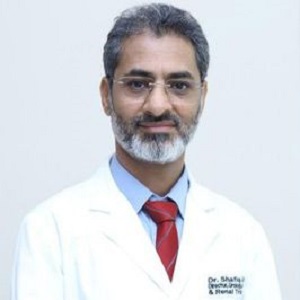
- Director – Urology, Andrology & Renal Transplants
- 16+ years
Profile Highlights:
Profile Snapshot of Dr. Shafiq Ahmed
- With over 16 years of experience, Dr. Shafiq Ahmed is a highly skilled urologist and robotic surgeon.
- He has previously worked at some of the best hospitals in India and was instrumental in establishing Renal Transplant Programmes in some of the top hospitals within Delhi-NCR.
- Dr. Ahmed also holds US credentials as a robotic uro-oncologist.
- He has authored numerous book chapters and articles in medical journals on urological diseases.
- As one of the leading urologists, robotic surgeons, and renal transplant surgeons, Dr. Ahmed has trained many upcoming doctors in his field.
- He has also been a prominent speaker at various medical conferences.
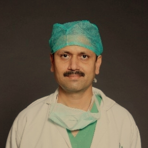
- Surgical Oncologist, New Delhi, India
- Over 20 years’ experience
Profile Highlights:
- Dr. S M Shuaib Zaidi is a Senior Consultant of Surgical Oncology at Apollo Cancer Institute of Indraprastha Apollo Hospital.
- His expertise lies in the surgical treatment of cancer of the lungs, esophagus, breast, GI tract, and mediastinal regions.
- Dr. Shuaib Zaidi has close to 2 decades of experience in the field of Surgical Oncology and his expertise lies in Thoracic Oncology surgery.
- Dr. Shuaib is known to have developed an innovative technique known as Radical Neck dissection for head and neck cancer surgery that helps in preserving the marginal mandibular nerve in order to ease the cosmetic outcome of the procedure.
- Dr. Zaidi also has experience in Robotic Surgery and received training in the procedure from IRCAD in Strass Bourg, France.
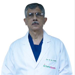
- Pediatric Cardiac Surgeon, New Delhi, India
- Over 40 years’ experience
Profile Highlights:
- Dr. Krishna Subramony Iyer is one of the best Pediatric Cardiac surgeons in India and specializes in congenital heart diseases.
- He has been practicing pediatric cardiac surgery for over 4 decades and has performed more than 10,000 surgeries through various procedures like double switch operation TAPVC repairs, Fontan and Fontan, arterial switch, and DORV.
- Dr. Iyer has been associated with Escorts Heart Institute for a long time and is responsible for establishing the first pediatric cardiac care program in North India in 1995.
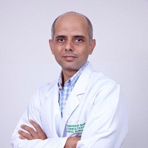
- Plastic & Cosmetic Surgeon, Gurugram, India
- Over 20 years’ experience
Profile Highlights:
- Dr. Adhishwar Sharma is a reputed plastic surgeon, with an extensive experience in plastic and reconstructive surgery.
- He has been associated with various hospitals like Fortis hospital Noida, Batra Hospital, Metro hospital Faridabad, and Central hospital Faridabad.
- He has an interest in Brachial Plexus reconstruction and lymphedema. He has published articles in various international and national journals. He is a member of various professional bodies.
- Endocrinologist, Diabetologist, New Delhi, India
- Over 26 years’ experience
Profile Highlights:
- Dr. Mohammada Asim Siddiqui is one of the best endocrinologists in India. He has an experience of over 26 years in the field. Dr. Siddiqui is active as a senior consultant – endocrinologist with Apollo Centre for Obesity, Diabetes, & Endocrinology, New Delhi.
- Dr. Siddiqui specializes in Internal Medicine & manages Diabetes, Endocrine and Metabolic Disorders, skin allergies, and Piles Treatment.
- His interest in Diabetes mainly focuses on young and gestational diabetes & Post-transplant & post-metabolic surgery diabetes.
- Dr. Siddiqui is also involved in various clinical research and has penned more than 50 articles in national and international journals.
- Robotic Urologist, New Delhi, India
- Over 27 years’ experience
Profile Highlights:
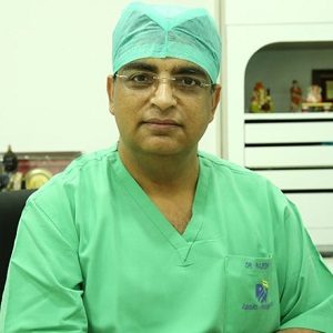
- Dr. Rajesh Taneja is one of the best Urologists in India, having experience of more than 27 years in the specialization. He is practicing at Apollo Hospitals, New Delhi as a senior consultant.
- Dr. Taneja specializes in surgeries to correct Congenital anomalies of urinary organs; Phimosis, PUJ Obstruction, Megaureter, Hydronephrosis, Vesicoureteric reflux (VUR), Hypospadias, Posterior Urethral Valves (PUV), Ectopia vesicae, and others.
- He is formally trained in the Robotic Surgical system and is one of the few urologists performing Robotic urological surgeries in India.
- Dr. Taneja has expertise in the Holmium Laser Enucleation of prostate technique.
- He has written numerous papers in national and international scientific publications.
- Dr. Rajesh Taneja also authored a book titled ‘Interstitial Cystitis’.
Best Hospitals in India for AVM Brain Treatment
Paras Hospital, Gurugram
- City: Gurugram, India
Hospital Highlights:
- Paras hospital was established in 2006 and is the 250 bedded flagship hospital of Paras Healthcare.
- The is supported by a team of doctors of international and national repute.
- The hospital is NABH accredited and also the first hospital in the region to have a NABL accredited laboratory.
- The hospital provides specialty medical services in around 55 departments including Neurosciences, Joint Replacement, Mother & Child Care, Minimal Invasive Surgery, Gynecology and Obstetrics, Ophthalmology, Dermatology, Endocrinology, Rheumatology, Cosmetic and Plastic surgery.
- The hospital is equipped with state-of-the-art technologies.
S L Raheja Hospital, Mahim, Mumbai
- City: Mumbai, India
Hospital Highlights:
- SL Raheja hospital is a 140-bed multi-specialty tertiary care hospital that is being managed by Fortis Healthcare Ltd.
- The hospital is a benchmark in healthcare and medical facilities in the neighborhood of Mahim & the western suburbs.
- L.Raheja Hospital, Mahim has one of the most effective ICU and Casualty care services.
- The hospital provides specialty medical services in Cardiology, Oncology, Neurology, Orthopedics, Mother & Child Care, and in Diabetes.
Wockhardt Hospitals, Mumbai
- City: Mumbai, India
Hospital Highlights:
- Wockhardt Hospitals were established in the year 1973, originally called First Hospitals and Heart Institute.
- Wockhardt Hospitals are super specialty health care networks in India, nurtured by Wockhardt Ltd, India’s 5th largest Pharmaceutical and Healthcare company.
- Wockhardt Hospitals is associated with Partners Harvard Medical International, an international arm of Harvard Medical School, USA.
- Wockhardt Heart Hospital performed India’s first endoscopic heart surgery.
- The hospital has a state-of-the-art infrastructure equipped with the latest technologies and modern equipment.
- It has special Centers of Excellence dedicated to the major specialties to provide hassle-free and high-quality clinical care.
Pushpawati Singhania Hospital & Research Institute, New Delhi
- City: New Delhi, India
Hospital Highlights:
- Established in 1996, Pushpawati Singhania Research Institute is one of the top hospitals in the NCR region, as well as one of the top facilities in India for gastroenterology. The hospital is one of South Asia’s first institutes in medical and surgical treatment for diseases related to digestion.
- The hospital is equipped with state-of-the art facilities coupled with the latest equipment as well as renowned consultants from various parts of India as well as other parts of the world.
Indian Spinal Injuries Center, New Delhi, India
- City: New Delhi, India
Hospital Highlights:
- The Indian Spinal Injuries Center (ISIC), provides state-of-the-art facilities for the management of all types of spinal ailments.
- Staffed with internationally trained, acclaimed, and dedicated spine surgeons, the hospital provides cutting-edge medical & surgical technology. The hospital provides comprehensive management of spinal injury, back pain, spinal deformities, tumors, osteoporosis, etc.
- The hospital performs motion-preserving spine surgeries including disc replacement and dynamic fixation, and minimally invasive spine surgeries such as endoscopic disc excision.
- The orthopedic service of the hospital covers all orthopedic ailments including trauma, joint diseases & replacements, oncology, pediatric orthopedics & upper limb ailment.
W Pratiksha Hospital, Gurgaon
- City: Gurugram, India
Hospital Highlights:
- W Pratiksha Hospital, Gurugram, is one of the best hospitals in the NCR region. It is also a top hospital in India for IVF. Since its inception, the hospital has performed over 5500 successful IVFs. The hospital also specializes in gynecology.
- With over 20 years of experience in providing quality healthcare, the hospital is known as one of the most trusted and valued health providers in India.
- Equipped with world-class medical facilities and advanced technology, the hospital’s doctors and clinicians also have a track record of delivering excellent results. The hospital is also known for focusing on preventive well-being as much as on curative treatment.
- The hospital has earned the trust of its patients, by providing the best available treatments at affordable costs.
Narayana Superspeciality Hospital, Gurugram
- City: Gurugram, India
Hospital Highlights:
- Situated near DLF Cyber City, Gurugram, Narayana Superspecialty Hospital is one of the top medical facilities in the Delhi NCR region, catering to the needs of the people. Known for its commitment to quality medical care and patient service, the hospital is a state-of-the-art facility with planned and well-equipped sections, which includes a spacious OPD area as well as comfortable patient rooms.
- It is the closest super-specialty hospital from Indira Gandhi International Airport towards Gurugram, and also the nearest super specialty hospital from DLF Cyber City. It is also close to major residential areas in Gurugram.
- It is part of the renowned Narayana Health Group. Established in 2000, by Dr. Devi Shetty, a renowned cardiac surgeon, it has grown to be one fo India’s leading healthcare groups.
Sir Ganga Ram Hospital, New Delhi
- City: New Delhi, India
Hospital Highlights:
- Sir Ganga Ram Hospital, New Delhi is known to provide the latest medical procedures with the latest technology in all of its units.
- The hospital has a team of reputed doctors, nurses, and healthcare professionals that ensure that patients receive quality care at affordable costs.
- Staffed with a team of highly qualified doctors, dedicated nurses, and paramedical and non-medical staff, the hospital aims to lead in healthcare delivery, medical education, training, and research.
- As per the vision of the founder, the hospital also provides free treatment to the economically weaker sections of society.
- Sir Ganga Ram Hospital also provides training to young doctors under the Diplomate in National Board(DNB) program. The DNB program at the hospital was started in 1984 and it is known for currently running the maximum number of DNB specialties in the country. It also has the distinction of having the first bone bank in India.
KIMS Hospital, Hyderabad
- City: Hyderabad, India
Hospital Highlights:
- KIMS Hospital (a brand name of Krishna Institute of Medical Sciences) is one of the largest and best multi-speciality hospitals in Hyderabad. The hospital provides various treatments to an enormous number of patients.
- The hospital has a capacity of more than 3000 beds. KIMS Hospitals offers different healthcare services in more than 25 specialities and super specialities.
- The hospital is equipped with modern medical equipment and technology. It has robotic equipment to provide minimal invasive techniques for patients.
- The hospital is aimed at providing world-class healthcare facilities and services at an affordable cost for patients.
- The various specialities and departments of the hospital include neurosciences, gastroenterology & hepatology, robotic science, reproductive sciences, dental science, oncological sciences, organ transplantation, heart and lung transplantation and mother and child care.
Fortis Hospital, Shalimar Bagh
- City: New Delhi, India
Hospital Highlights:
- Fortis Hospital in Shalimar Bagh is a multi-super specialty hospital that strives to provide world-class patient care by leaving no stone unturned.
- Fortis, Shalimar Bagh, with 262 beds and a 7.34-acre footprint, provides the best level of medical care through its team of doctors, nurses, technicians, and management professionals.
WHAT IS AN AVM BRAIN?
What causes Brain AVM?
The primary cause of AVM is still unclear. Some people have brain AVM by birth while some others develop the same later. A notable point is the passing down of this malformation amongst families, genetically.
Occurrence rate of AVMs
Brain AVMs occur very rarely and their occurrence rate is around 1% of the population. However, you might observe male predilection for AVM of the brain.
Where AVMs occur
Although an AVM may develop anywhere within the body, the most common sites are the brain and spine. However, brain AVM is rare and affects only a small chunk of the human population.
Symptoms of Brain AVMs
You may not notice any symptoms of brain AVM till it ruptures. Rupturing of AVM of the brain may result in bleeding within the brain which is termed hemorrhage. A majority of the patients with brain AVM experience hemorrhage as the first sign. Other symptoms of brain AVM include:
- Constant pain in any specific area of the head
- Problems with speech
- Progressive loss of neurological function
- Numbness or paralysis
- Dizziness
- A problem in understanding language
- Nausea and vomiting
- A problem in performing certain tasks that require planning
- Back pain
- Loss of coordination
- Seizures
- Muscle weakness in a specific part of the body
- Loss of consciousness
- Weak lower limbs
- Confusion
- Vision loss
- Serious unsteadiness
- Hallucinations
- Inability to understand what others are saying
- Loss of control on eye movements
- Tingling sensations
Causes of bleeding AVM
The abnormal blood vessels in brain AVM are weak. Hence, they direct blood in a direction against the tissues of the brain. As there is dilation of the abnormal blood vessels with time, they tend to burst. A brain AVM ruptures and bleeds because of the blood flow in a high pressure coming from the arteries.
Chances of AVM bleeding
A brain AVM generally develops between the age of 10 years to 40 years. On average, the chances of bleeding of brain AVM is between 1% to 3% per year. If it bleeds for the first time, the patients are at risk of recurrent bleeding within a short period. Teenagers and the young age group people are at a high risk of experiencing recurrent bleeding.
When to visit the doctor?
If you notice any signs or symptoms of brain AVM, you must visit the doctor immediately. These cases require emergency medical attention otherwise they may bleed profusely & prove to be fatal.
Stages of brain AVM
- Stage 1 of Quiescence: AVM brain malformation is quiet in this stage. The skin surface above the AVM is red and feels a little warm.
- Stage 2 of Expansion: The malformation increases in size to become larger and it is easy to feel the pulse within the malformation.
- Stage 3 of Destruction: The brain AVM begins to bleed and pain.
- Stage 4 of Decompensation: This stage may turn out to be fatal as heart failure occurs in most cases.
Can AVM bleeding prove fatal?
Around 10% to 15% of deaths occur after each AVM brain bleeding. Some patients experience permanent brain damage that accounts for around 30% of the total cases. Every bleed contributes to the damage of normal tissues within the brain that may alter the normal brain functions temporarily or permanently.
Types of Brain AVM
There are 5 different types of brain AVM. These types are listed as under:
- True AVM: This is the most common type of malformation in which the abnormal blood vessels tangle. However, there is no interfering normal tissue of the brain.
- Venous malformation: This malformation involves only the abnormal veins.
- Dural fistula: Dura mater, the covering of the brain, often gets involved in an abnormal connection with the blood vessels. This results in the formation of a dural fistula. There are three types of dural fistulas namely transverse- sigmoid sinus dural fistula, dural carotid-cavernous sinus fistula, and sagittal sinus-scalp dural fistula.
- Cryptic or occult AVM or cavernous malformations: This type of vascular brain malformation doesn’t draw away large quantities of blood. Such malformations may cause seizures and bleed.
- Hemangioma: Abnormal blood vessels at the brain or skin surface are termed hemangioma.
Risk factors of Brain AVM
Males are at a higher risk of developing brain AVM. Apart from this, families with a history of brain AVM have generic passing down of the same.
Complications of Brain AVM
The complications of brain AVM include stroke, speech or movement problems, low quality of life, developmental delays (in children), and numbness in specific parts of the body. Other complications associated may be:
- Weaker blood vessels: An AVM brain exerts pressure on the weaker blood vessels. This may result in an aneurysm or a bulge in the walls of the blood vessels that may rupture later.
- Hemorrhage: AVM causes the walls of the affected veins and arteries to weaken which results in bleeding within the brain or hemorrhage. While hemorrhage goes undetected in some people, some others experience episodes that may be life-threatening.
- Brain damage: In many cases, AVM increases in size to constrict some portions of the brain. Because of this, protective fluids are unable to circulate freely and instead collect at a place. This may cause brain tissue to get pushed against the skull resulting in brain damage.
- Reduced oxygen supply: When brain AVM develops, blood circulates directly to the veins & the arteries. Because of this, surrounding tissues are unable to absorb oxygen from the blood, and these tissues weaken or die.
How Brain AVMs are diagnosed
Your doctor will review all your symptoms and perform a thorough examination. For forming a diagnosis, he or she might ask you to undergo certain tests. These tests are usually performed by neuroradiologists. They are professionally trained to conduct imaging tests for finding the cause and the diagnosis of the condition.
Tests
- CT scan: A Computerized Tomography scan creates a cross-sectional radiographic image of the brain through an x-ray series. Sometimes, doctors perform angiograms which involves injecting a dye. This dye is injected into the veins through a tube so that minute details can be seen.
- Cerebral arteriography: Also called cerebral angiography, it helps to accurately diagnose AVM. It reveals not only the characteristics of arteries that supply AVM and the draining veins but also the exact location. These details are essential for the diagnosis and treatment of the condition. Your doctor will insert a tube called a catheter in the artery of the groin region. He or she will use x-ray imaging to thread it to the brain. Injecting the dye will make the blood vessels clear enough to be seen in the x-ray imaging.
- MRI: Magnetic Resonance Imaging uses radio waves and magnets to obtain precise brain images. This test will provide you with the exact location of AVM within the brain along with any other bleeding spot. Alternatively, your doctor might perform Magnetic Resonance Angiogram using the dye.
Treatment of Aterivenous Malformations (AVMs) of Brain
There are ample treatment options for this malformation. The primary goal is to avoid hemorrhage but it is vital to consider the neurological complications. Your health, gender, age, location, and size of the blood vessels are some critical factors that help in determining the right treatment option.
Stereotactic radiosurgery
This type of treatment involves radiation for the destruction of the brain AVM. To simply put, this isn’t surgery but involves targeting of AVM by the radiation beams. This damages the abnormal blood vessels leaving a scar. The scarred blood vessels clot off within a few years of the treatment. If you have a small AVM that can’t be removed with conventional surgery then your doctor might suggest SRS. People having hemorrhages that may turn fatal also undergo SRS treatment
Endovascular Embolization
It is also called interventional neuroradiology. Your doctor will insert a catheter tube into the artery of your leg and thread it with the brain blood vessels. The catheter is placed in one of the arteries that feed AVM followed by injecting of the embolizing agent. This will block the blood flow to the feeding artery. This is a minimally invasive procedure and is usually done before any surgery. It reduces the chances of AVM bleeding and reduces its size. Endovascular embolization also decreases the chances of stroke-type symptoms.
Surgical removal or resection
If the AVM is bleeding or is located in an easily accessible area of the brain, resection via conventional brain surgery is a preferable treatment option. Your doctor will temporarily remove a portion of your skull to perform resection. AVM can be easily removed with the help of special clips followed by reattachment of the skull bone. Suturing of the incision follows this process to close the scalp region.
Medical therapy
If you aren’t experiencing any symptoms of AVM or the AVM is located in an inaccessible area, you will undergo conservative management. Your doctor will ask you to avoid excess workouts and exercise. Also, you will have to discontinue the intake of blood thinners or anticoagulants like warfarin.
Specialists for brain AVM treatment
A brain AVM treatment is performed by specialists who hold a specialization degree in such cases. They may be:
- A neurosurgeon or radiation therapist who performs stereotactic radiosurgery treatment.
- A stroke neurologist who is proficient in the medical management of such malformations. They can also diagnose easily and perform imaging of the head, neck, and brain region.
- The vascular neurosurgeon can perform surgical removal of AVM of the brain.
- An endovascular neurosurgeon or interventional neuroradiologist who excels in endovascular therapy.
Prevention and prognosis
Most commonly, an AVM occurs by birth or sometime later after the birth of the child. Prevention is difficult as the causes are unknown for a majority of the cases. However, the best thing would be to seek medical attention as soon as you notice the first symptom for a better prognosis. Timely medical treatment always helps in easy management and provides a better quality of life. People who get to know about AVM through bleeding usually die. Some others experience seizures and problems with the nervous system. Many people with undetected AVM till their 50s or 60s experience no symptoms and are stable.
Follow up
You need to visit your doctor regularly for follow-ups after getting surgically operated on for brain AVM. Your doctor might advise you imaging tests again to ensure there is no recurrence and the malformation has resolved completely.
FAQs on Brain AVM
Brain AVM is curable within 2-3 years of treatment. Radiosurgery is often a treatment choice for AVMs that are small in size but in exceptional cases, one may opt for it even for the removal of large AVMs.
One in 2000 people develop brain AVM. A majority of the people live normally with it but they have a risk of bleeding AVM at some time. Sometimes, they may also suffer a stroke due to AVM.
In most cases, AVMs do not rupture. However, there may be recurrent bleeding after it ruptures once.
Most of the asymptomatic AVM cases have a history of bleeding. Due to increased stress, you may have high BP. This may cause the AVM to bleed as the blood will flow at a fast rate and the vessel walls will be weak.
The most important thing to keep in mind is that you must avoid anything that elevates the blood pressure. If you perform a high workout or a strenuous activity, your blood pressure may rise. This will result in strain on your malformation and there are high chances of AVM rupture or bleeding.
AVM is a collection of abnormal blood vessels that restrict blood circulation to the brain. It may sometimes be accompanied by vascular tumors that are surgically removed.
Although there are no cases of passing down of AVM in families, a mere 5% of cases are due to autosomal dominance. However, the causes of AVM are still unknown in a majority of the cases as it is present by birth.
A Grade 4 or Grade 5 AVM is considered a large AVM as it is big and deep within the brain. In most cases, a large AVM is considered to be a risky malformation to be operated upon.
No, AVM hasn’t been identified as a condition of disability but severe complications of its rupture may result in the disability of the person.
No, a brain AVM does not cause any kind of personality changes. However, a patient may experience emotional changes due to the stress of the same.
The warning signs of a brain aneurysm are seizures, headache, nausea, vomiting, loss of consciousness, stiff neck, drooping eyelids, and blur vision.
The symptoms of a brain aneurysm headache are pulsing sensation and throbbing pain. This may last for a couple of hours to a few days and it may be debilitating. The patient might also experience sensitivity to extreme sound and sunlight.
Medical therapy can help you get relief from the symptoms of AVM like headache and pain. You can perform routine activities only after several weeks following the surgery. Most patients require around 4 to 6 months to completely recover from the condition.
There will be a headache suddenly that is severe enough to disturb the patient. The patient will experience headaches in only a specific region of the head with a ringing sound in the ear.
A Cavernoma or a cavernous angioma or a cavernous malformation is one of the types of AVM of the brain. It also contains a collection of weakened blood vessels that block the blood flow.

