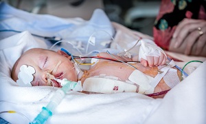
Spina Bifida
Spina Bifida also known as neural tube defect, is a birth defect that occurs when your spine and spinal cord don’t form correctly. The neural tube is the structure in a developing embryo which eventually becomes the brain of the child, spinal cord as well as the tissues enclosing them.
In normal cases, the neural tube forms early during pregnancy and it closes by the 28th day after conception. In babies with spina bifida, one portion of the neural tube fails to close or develop properly, which results in defects in the spinal cord and the spinal bones.
Spine Bifida can be mild as well as severe, as it depends on the type of defect, location, size as well as complications. When required, early treatment for spina bifida will involve surgery, but the problem isn’t always resolved by such treatment.
Types of Spina Bifida
There are three main types of spina bifida, which include myelomeningocele, meningocele and spina bifida occulta.
1.Myelomeningocele- The most common and serious type of spina bifida which involves a sack outside the opening in the back of the baby somewhere in the spine, which contains parts of the spinal cord as well as nerves which will get damaged. People who have myelomeningocele can have physical disabilities which may be moderate to severe and some of such disabilities can include:
- Incontinence
- Difficulty in going to the washroom
- Inability in moving or feeling one’s legs or feet
2.Meningocele- Meningocele is another type of spina bifida which involves a sack of fluid outside an opening in the back of the baby. However, the sack doesn’t contain any part of the spinal cord, as there isn’t much nerve damage, this kind of spina bifida only leads to minor problems.
3.Spina Bifida Occulta- This is another mild type of spina bifida which also goes by the term ‘hidden spina bifida’, as it doesn’t cause any disabilities and might go unnoticed until later in life. Usually, there is no opening the in back of the baby but only a gap in the spine. In this type, there is also damage to the spinal cord or the nerves.
Symptoms of Spina Bifida
Each type of spina bifida shows different symptoms and they can vary from person to person as well.
Symptoms of myelomeningocele spina bifida can include:
- Over some of the vertebrae the spinal canal is open, often in the middle or lower part of the back
- Weak or paralyzed muscles in the leg
- Membranes and spinal cord pushed outside the back in a skin-covered sack
- Deformed feet
- Uneven hips
- Seizures
- Scoliosis
- Issues with the bowel and bladder
Symptoms of meningocele can include the following:
- Sack that is visible at birth
- Small opening at the back
- Membranes which are pushing out through the opening in the vertebrae into the sack
- Normal spinal cord development
Spina bifida occulta symptoms include- - No visible opening outside
- A gap in between vertebrae
- No fluid-filled sack outside the body
- Small cluster of hair on the back
- A small area of extra fat on the back
- Small birthmark such as a dimple on the back
Causes of Spina Bifida
All the exact causes of this ailment are still not understood properly. However, it is known that combination of genetics and environmental factors are involved. A child who is born with spina bifida might not have any relatives with this condition but genetics does play a factor. It is also believed that lack of folic acid (vitamin B-9) can play a role in this ailment. Other factors that are said to play a role include obesity, some kind of medications and uncontrolled diabetes in the mother.
Diagnosis of Spina Bifida
Pregnant women will be offered prenatal screening tests for checking for spina bifida and other birth defects. These tests aren’t perfect. Some mothers had positive blood tests have babies without spina bifida. Even if the result is negative, there is a slight chance that the spina bifida is present, so consider talking to your doctor regarding prenatal testing, its risks and you now need to handle the results.
Though spina bifida can be screened using maternal blood tests, typically ultrasound is used for diagnosis in most cases.
Maternal Serum Alpha-Fetoprotein test
For Maternal Serum Alpha-Fetoprotein test, a sample of the mother’s blood is drawn to be tested for alpha-fetoprotein, a protein that is produced by the baby. It’s quite normal for a little amount of AFP to cross the placenta and enter the mother’s bloodstream, but abnormally high levels of AFP suggests that the baby has a neural tube defect, which can be spina bifida, even though high levels of AFP don’t occur always in spina bifida.
Test to Confirm high AFP levels
Varying levels of AFP can also be caused by other factors, which include a miscalculation in the fetal age of multiple babies. Therefore, your doctor might order another blood test for confirmation, If the results are still high, you will need even further evaluation, which will include an ultrasound exam.
Other blood tests
Ultrasound
Fetal ultrasound is the most accurate method for diagnosing spina bifida in the baby before delivery. Ultrasound may be performed during the first trimester and second trimester. Spina bifida can be diagnosed accurately in the second-trimester ultrasound scan. This exam, therefore, becomes quite important to identify and rule out congenital anomalies like spina bifida.
An advanced ultrasound can also detect signs of spina bifida, which includes an open spine or particular features in the brain of the baby, which indicate spina bifida. An expert can easily assess the severity of the ailment using ultrasound.
Amniocentesis
If the prenatal ultrasound confirms the diagnosis of spina bifida, your doctor might request amniocentesis. During this process, your doctor removes a sample of fluid from the amniotic sac surrounding the baby using a needle. The purpose of this examination is to rule out genetic diseases, even though spina bifida is rarely associated with genetic diseases.
Treatment options for Spina Bifida
The treatment for spina bifida may vary as it always depends on the severity of the condition. While spina bifida occulta won’t require any treatment, other types do.
Surgery before birth
Nerve function in babies who have spina bifida can get worse after birth, if spina bifida isn’t treated. Prenatal surgery to treat spina bifida takes place before the 26th week of pregnancy. In this procedure, surgeons expose the uterus in the pregnant mother surgically open the uterus and repair the baby’s spinal cord. In some patients, this procedure can be performed even less invasively with a fetoscope through ports in the uterus.
According to research, children with spina bifida who goes through fetal surgery have reduced disability and are less likely to require crutches or any such walking devices. Fetal surgery can reduce the risk of hydrocephalus as well. Consult with your doctor whether this procedure is appropriate for you. Talk regarding the potential benefits and risks such as possible premature delivery as well as other such complications.
It is also quite important to have a comprehensive evaluation to determine whether fetal surgery is feasible. This specialized surgery should only be done at a proper health-care facility that has experienced doctors in fetal surgery. A multispeciality team approach and neonatal intensive care are also important. The team typically includes a fetal surgeon, a pediatric neurosurgeon, a maternal-fetal medicine specialist, a fetal cardiologist as well as a neonatologist.
Cesarean birth
Surgery after birth
Surgery is required for babies with myelomeningocele, and the earlier it is performed, the earlier the risk of infections will be minimized. It will also help in protecting the spinal cord from being exposed to further trauma.
During this procedure, the neurosurgeon will place the spinal cord along with the exposed tissue inside the baby’s body, after which he will cover them with muscle and skin. At the same time, the neurosurgeon might place a shunt in the baby’s brain for controlling hydrocephalus.
Treatment for complications
For babies having myelomeningocele, there has been already irreparable nerve damage and usually a multispeciality team of surgeons, physicians and therapists might be required. Babies having this ailment might need even more surgery to treat a variety of complications. Some of these complications might include weak legs, bladder and bowel problems or hydrocephalus, which usually begins soon after birth.
Depending on the severity of spina bifida and the complications, the treatment options can include:
- Walking and mobility aid- Some babies can start exercises to prepare their legs for walking with the aid of braces of crutches as they get older. Some children might need walkers or wheelchairs. Mobility aids combined with regular physical therapy can help a child in becoming independent. Even children who require wheelchairs can learn to function quite well and become self-sufficient.
- Bowel and bladder management- Routine evaluation of bowel and bladder evaluations as well as management plans can help in the reduction of the risk of organ damage and illness. Evaluations can include X-rays, kidney scans, blood tests, ultrasounds and bladder function studies. These evaluations will have to be done frequently in the first few years of life but it can become lesser, as the child continues to grow.
- Surgery for hydrocephalus- Most babies who have myelomeningocele, will require a surgically placed tube which can allow fluid in the brain to drain into the abdomen. This tube might be placed just after the baby is born, during the surgery to close the sac located on the lower back or later as the fluid accumulates. A less invasive option is also available which is called endoscopic third ventriculostomy. However, candidates are chosen very carefully for this and they need to meet certain criteria. During this procedure, the surgeon will use a small video camera to see inside the brain. He makes a hole in the bottom of between the ventricles so cerebrospinal fluid may flow out of the brain.
- Treatment and Management of other complications- Special equipment which includes bath chairs, commode chairs as well as standing frames might help with daily activities. Whatever the issue, orthopedic complications or tethered spinal cord, most of them can be treated or managed to help improve the child’s quality of life.
Prevention of Spina Bifida
Taking folic acid in supplement form at least one month before one’s conception and continuing through the first trimester of the pregnancy can greatly reduce the risk of spina bifida and other neural tube defects. Having plenty of folic acid in the system by the early weeks of pregnancy is important if you want to prevent spina bifida. Since many women don’t realize that they are pregnant till this time, it is recommended by experts that women who are of childbearing age, should take at least a daily supplement of 400 micrograms of folic acid. Food such as rice, pasta, enriched bread and some breakfast cereals are rich in folic acid.
Women who are planning or expecting pregnancy should also remember to consume at least 400 to 800 mcg of folic acid per day. The human body doesn’t absorb folate as easily as it can absorb synthetic folic acid and therefore it is important that you take vitamin supplements as well. It is also quite possible that folic acid will help to reduce the risk of other birth defects, including cleft lip, cleft palate as well as some congenital heart defects.
It is quite a good idea to eat a healthy diet, including foods that are rich in folate or enriched with folic acid. It is seen to be present naturally in several foods, such as egg yolks, milk, beans, peas, citrus fruits, juices, avocados, dark green vegetables, etc.


