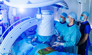
Hip arthroscopy
Arthroscopy is the examination of the interior of a joint by using an arthroscope, a flexible, small fiber-optic tube with a small camera connected to a monitor. This can allow a surgeon to get a magnified view of your joint. Specially designed arthroscopic surgical tools are also used to perform various types of minimally invasive joint surgery. Hip arthroscopy, which is known as a ‘hip-scope` is a minimally invasive procedure, in which an orthopedic surgeon examines the hip joint with the help of an arthroscope.
This procedure can help the surgeon diagnose the cause of hip pain or any other problem in your joint. Some hip conditions might also be treated arthroscopically. For this kind of procedure, the surgeon needs to make a few additional small incisions to create access points for various arthroscopic needles, scalpels, or other special surgical tools.
Purpose
Hip arthroscopy can be recommended by your doctor if you are having a painful condition that doesn’t respond well to nonsurgical treatment. Nonsurgical treatment generally includes rest, medications, and physical therapy or injections that can reduce the inflammation.
Hip arthroscopy can help to relieve painful symptoms of several problems, that can damage the labrum, articular cartilage, or any other soft tissues that are surrounding the joint. This damage can result from an injury. In some cases, other orthopedic conditions can also result in problems, such as:
- Femoroacetabular impingement (FAI) is a disorder in which extra bone develops along the acetabulum or on the femoral head. This bone overgrowth, which is known as spurs, cause damage to the soft tissues of the hip during movement. Sometimes bone spurs can develop in both the acetabulum and femoral head.
- Snapping hip syndromes cause a tendon to rub across the outside of the joint. This type of snapping or popping is generally harmless and does not require treatment. The tendon in some cases can get damaged due to repeated rubbing.
- Dysplasia is another condition in which your hip socket becomes abnormally shallow. This can put more stress on the labrum for keeping the femoral head within the socket, and this makes the labrum more susceptible to tearing.
- Loose bodies are fragments of bone or cartilage that become loose and move around within the joint.
- Synovitis can cause inflammation in the tissues that surround the joint to become inflamed.
- Hip joint infection
Preparation
It is likely that your orthopedic surgeon will recommend you to see your primary doctor so that he/she is able to assess your general health prior to the surgery. He/she will identify any problems that can interfere with the procedure. If you are having certain health risks, a more extensive evaluation might be required before the surgery.
If your physical condition is healthy, then it is likely that your hip arthroscopy will be performed as an outpatient procedure. This means that you won’t need to stay at the hospital overnight.
Make sure that you inform your surgeon regarding any medications or supplements that you might be taking. You might need to stop taking some of these before the surgery.
The hospital or surgery center will contact you ahead of time to provide specific details of the procedure. Make sure that you follow the instructions regarding when you need to arrive, as well as when as you need stop consuming food and drink prior to your procedure.
Procedure
At the start of your procedure, your leg is going to be put in traction. This means that your hip is going to be pulled away from the socket enough for your surgeon to insert instruments, see the entire joint, and perform any treatment as needed.
Surgeons generally draw lines on the hip to indicate specific anatomy structures, such as bone, nerves, and blood vessels, as well as incision placements and portals for the arthroscope.
After the traction is applied, your surgeon will be making a small puncture in your hip, which will be about the size of a buttonhole, for the arthroscope. Through the arthroscope, your surgeon is able to view the inside of your hip and identify any damage.
In the meantime, fluid flows through the arthroscope, to keep the view as well as to control any bleeding. Images from the arthroscope are projected on the video screen so that your surgeon is able to see the inside of your hip as well as any problems. Next, your surgeon is able to evaluate the joint before he/she begins any specific treatments.
Once your surgeon is able to properly identify the problem, he/she will insert small instruments through separate incisions for repairing it. There are various procedures that can be done, depending on your needs.
For tasks such as shaving, cutting, suture passing and grasping, and knot tying, the surgeon uses specialized instruments. In most cases, special devices are used for anchoring stitches into the bone.
The length of the procedure generally depends on what your surgeon finds and the amount of work that needs to be done. At the end of the surgery, the incisions are generally stitched or covered with skin tapes. An absorbent dressing is next applied to the hip.
Recovery
Symptoms usually improve immediately following the procedure, but sometimes as the irritated joint lining heals, there might be a recurrence of some pain.
You might also feel a watery sensation in the hip or might even hear gurgling noises resulting from the fluid used during surgery. However, this will be absorbed by the body quickly. Swelling should subside in around a week, and any sutures will be removed in seven to ten days. Depending on the specific method used during your procedure, your full recovery time is going to vary.
Generally, hip arthroscopy patients need to use crutches for a week or two, after surgery, and they also require around six weeks of physical therapy. After around three to six months, patients should experience no pain after any physical activity.
Risks & complications
Generally, complications from hip arthroscopy are uncommon.
Any surgery in the hip joint has a small risk of injury to the surrounding nerves or blood vessels. Injury is a possibility in the joint itself.
There is also a small risk of infection, as well as blood clots forming in the legs.


