Best Doctors in India for Myocardial Perfusion Imaging
- Interventional Cardiologist, Gurugram, India
- Over 10 years’ experience
Profile Highlights:
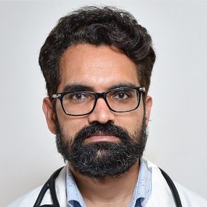
- Dr. Jagdeep Yadav is one of the best cardiac surgeons in Gurugram. He is particularly interested in non-coronary therapies such as peripheral interventions and device closures.
- Dr. Jagdeep Yadav is skilled in using modern techniques in interventional cardiology and non-invasive cardiology procedures. He employs cutting-edge technologies such as IVUS, OCT, FFR, and IVL to make life better in complex cardiac therapies.
- Cardiologist, Gurugram, India
- Over 15 years’ experience
Profile Highlights:
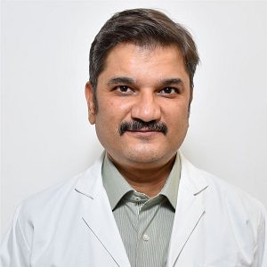
- Dr. Sanjat Chiwane is an MCI Certified young cardiologist in Gurugram.
- His career success measured 1200+ coronary angioplasties, 100 Balloon mitral valvuloplasties, and 100 renal angioplasties.
- Continuing with his meticulous career, the doctor also worked as a Clinical Instructor in Medicine and Cardiology at Mumbai University. He added Organ Transplantation to his expertise.
- Cardiologist, Gurugram, India
- Over 16 years’ experience
Profile Highlights:
- Dr. Kuldeep Arora is a promising cardiologist in Gurugram. He is a specialist in radial angiography and angioplasty.
- Dr. Kuldeep Arora has expertise in Interventional Cardiology and performed around 12,000 therapeutic cardiac interventions.
- He also assisted with the device closure of atrial septal abnormalities, Pacemaker implantations, ICD, and CRT, besides management of heart attacks.
- Cardiovascular Surgeon, Cardiac Surgeon, Gurugram, India
- Over 32 years’ experience
Profile Highlights:
- Dr. Vijay Kohli is one of the best cardiac surgeons in India. He has brought an evolution in the spectra of Cardiac Surgery, wherein, he was the first leading surgeon to perform CABG on a beating heart in Kathmandu, Nepal.
- Vijay Kohli performed the first coronary artery bypass surgery in Jammu Medical College in 2001.
- Additional Director Cardiology
- 15 Years Experience
Profile Highlights:
Profile Snapshot of Dr. Hemant Gandhi
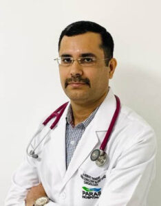
- Hemant Gandhi is a distinguished figure in the field of interventional cardiology with over 15+ years of extensive experience.
- He is recognized for his proficiency in handling cases involving congenital heart defects such as ASD and PDA, along with peripheral angiography, angioplasty, and balloon valvuloplasty.
- Gandhi holds the distinction of being the first D.M. (Cardiology) candidate to graduate from the prestigious PGIMER and Dr. RML Hospital, New Delhi.
- Beyond his clinical practice, he is actively engaged in the education and training of medical students in the specialized domain of Cardiology.
- His contributions to academia are further underscored by a significant number of publications that enrich the field with his insights and research findings.
Best Hospitals in India for Myocardial Perfusion Imaging
Myocardial Perfusion Imaging
Our heart muscle needs a steady flow of blood for functioning properly and staying healthy. Myocardial Perfusion Imaging (MPI) is a non-invasive method that lets your doctor evaluate this blood flow. The scans can also be used to look for damage after a heart attack and for determining if any previous treatment has helped.
While perfusion assessment might require you to exercise, the testing process is safe as well as painless.
Purpose
An MPI test is able to show how well blood is able to flow through the heart muscle. If the test shows a lack of blood flow during stress or exercise but is normal at rest, then it can mean that an artery that carries blood to your heart might be narrowed or blocked.
If the test is showing a lack of blood flow to a portion of the heart muscle during exercise or stress, and at rest, it can mean that your heart muscle is scarred, possible from a previous heart attack.
MPI tests can help your doctor in various ways such as:
- Finding out if there are blockages or narrowings in your coronary (heart) arteries if you are having chest discomfort
- Determining if you need to undergo a coronary angiogram
- Determining if you have heart damage from a heart attack if your heart is not functioning normally
- Deciding whether you would benefit from the coronary stent or bypass surgery for treating your chest discomfort or helping an abnormal pumping function go back to normal
- Determining how well your heart is able to handle physical activity
- Determining if a heart procedure you had to improve your blood flow is working properly
Preparation
Let your doctor know about any medications that you take, such as over-the-counter medicines, vitamins, or herbs. He/she may ask you not to take them before your test. Remember not to stop taking your medication until your doctor tells you to.
Your doctor might ask you to avoid certain foods, such as caffeine-containing beverages, such as tea, coffee or soft drinks, or chocolate, for around 24 hours before the test is performed. The test might need to be postponed or canceled if you consume caffeine.
Remember to wear comfortable and loose-fitting clothing as well as comfortable shoes to exercise in.
Procedure
The test is performed in a hospital or a clinic with special equipment.
The technician places small metal disks known as electrodes on your chest, arms, and legs. These disks have wires that hook up to a machine for recording your electrocardiogram. The ECG can keep track of your heartbeat during the test, and can be used to tell the camera when taking a picture is required.
You will next wear a cuff around your arm in order to keep track of your blood pressure. Your technician will be putting an intravenous line in your arm. Then you will need to exercise on an exercise bicycle or on a treadmill. If you are unable to exercise, due to certain conditions, then your IV line is connected to a bag that has a medicine, for increasing the blood flow, to your heart. This medicine has an effect similar to when you exercise, and makes the heart go faster.
When you reach the peak activity level, you will need to stop after which you will receive a small amount of radioactive material through your IV line.
You will lie still on your table for a few minutes while the gamma camera will take pictures of the heart. During this time, several scans are done to provide pictures of thin slices of your entire heart from all different angles. It is also very important to be completely still with your arms above your head, as the pictures are being taken.
While you are resting, you will be receiving more tracer, and another set of pictures will be taken as well. This set of images is going to be compared to the images taken after exercise or stress.
The test generally takes between 3- 4 hours. Some labs can choose to do the two parts of the test on different days.
After the procedure
You should be able to resume your normal routine right away. Remember to drink plenty of water to flush the radioactive material from your body.
Next, you will need to make an appointment with your doctor in order to discuss the test results and the next steps.
Risks
There are certain risks that are associated with an MPI test.
Sometimes the exercise part of the test can lead to rare instances of abnormal heart rhythms or chest pain. Sometimes it might also lead to a heart attack due to the stress on the heart that is caused by the exercise.
The needle that is used to inject the IV might cause some pain. The injection of the radioactive tracer might also cause a little discomfort. Although rare, sometimes there might be allergic reactions to the tracer.
Make sure you talk to your doctor regarding the amount of radiation used during the procedure and the risks related to your particular situation.
Depending on your specific medical conditions, there might be sometimes a few other risks as well. Therefore before you undergo the procedure, make sure your doctor knows about all your medical conditions.


